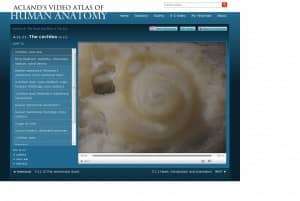
Wolters Kluwer Health has announced a new addition to Acland’s Video Atlas of Human Anatomy that it says sheds new light on the intricate and hard to ascertain anatomy of the inner ear. More than two years in the making, these new online educational videos are said to showcase the most innovative and revealing dissections of the human inner ear ever captured.

Dr Acland achieves this unique perspective using innovative camera rotation techniques that he pioneered with his colleagues at the University of Louisville School of Medicine. To make the new inner ear videos, Dr Acland used a high-speed micro drill, which creates a significant amount of bone dust. The only way to perform a clean dissection and present the results to the camera was to shoot underwater, which allows the dust to be carried away and helps support the delicate structures that would normally collapse in the open air. The resulting product reveals new insights and never-before-seen imagery of this complex part of the human body.
“There’s not a single book that allows for a three-dimensional understanding of the inner ear,” says Dr Acland, Professor Emeritus in the Department of Anatomical Sciences at the University of Louisville School of Medicine and creator of Acland’s Video Atlas of Human Anatomy. “That’s because its structures are intricately shaped, very small and delicate, and encased in rock-hard bone. Illustrators have struggled over two centuries to produce meaningful two-dimensional images of this very three-dimensional structure.”
With the advent of these new videos, students are able to view these structures three-dimensionally for the first time. Because the inner ear is so difficult to dissect, most students have never seen it in its true form. Many instructors, in fact, have struggled to understand it and as a result, many institutions have removed it from their curricula. Dr Acland himself purposely omitted the content from Acland’s Video Atlas when it was first developed due to the challenges it posed for him as a microsurgeon and videographer.
Dr Acland expects, however, that these videos will help reinvigorate study of inner ear anatomy by making it more accessible and less difficult to teach. Although it will benefit students in medical and related health sciences, even the most accomplished otolaryngologists will enjoy the series, says the publisher.
A total of five new instructional videos dedicated to the inner ear have been added. With a run time totaling more than 22 minutes, these clips cover the bony labyrinth and its relationships; the membranous labyrinth and fluids of the inner ear; the vestibule of the inner ear; the semicircular ducts; and the cochlea. Also included is a series of 3-D animations and numerous histology images to supplement understanding.
Acland’s Video Atlas of Human Anatomy contains 333 video clips of real human anatomic specimens in their natural colors. Organized into 5 volumes including the upper and lower extremities, the trunk, head and neck, as well as the internal organs, the Atlas shows moving structures—muscles, tendons, and joints—making the same movements that they make in real life. It is available in 6 or 12 month subscriptions to access either the full site or just the head and neck volume.
Source: Wolters Kluwer Health


